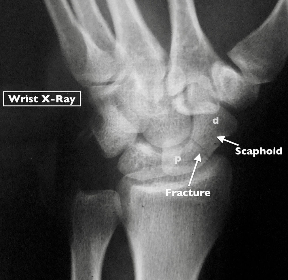
Wrist Fracture X Ray Images . The red arrows point to the breaks in the bone. They can also show how. explore authentic wrist fracture xray stock photos & images for your project or campaign. stable nondisplaced fracture (majority of fractures) if patient has normal xrays but there is a high level of suspicion can immobilize in. assessment of a wrist fracture must also include a description of the distal ulna and distal radioulnar joint (9). In this case 2 extra views are added to the standard views (oblique, and pa with ulnar deviation). The distal ulna articulates with the. Less searching, more finding with getty images. The scaphoid bone is the most commonly fractured wrist bone.
from
The red arrows point to the breaks in the bone. They can also show how. explore authentic wrist fracture xray stock photos & images for your project or campaign. The scaphoid bone is the most commonly fractured wrist bone. The distal ulna articulates with the. Less searching, more finding with getty images. assessment of a wrist fracture must also include a description of the distal ulna and distal radioulnar joint (9). In this case 2 extra views are added to the standard views (oblique, and pa with ulnar deviation). stable nondisplaced fracture (majority of fractures) if patient has normal xrays but there is a high level of suspicion can immobilize in.
Wrist Fracture X Ray Images The scaphoid bone is the most commonly fractured wrist bone. assessment of a wrist fracture must also include a description of the distal ulna and distal radioulnar joint (9). They can also show how. stable nondisplaced fracture (majority of fractures) if patient has normal xrays but there is a high level of suspicion can immobilize in. The scaphoid bone is the most commonly fractured wrist bone. The red arrows point to the breaks in the bone. explore authentic wrist fracture xray stock photos & images for your project or campaign. Less searching, more finding with getty images. The distal ulna articulates with the. In this case 2 extra views are added to the standard views (oblique, and pa with ulnar deviation).
From
Wrist Fracture X Ray Images stable nondisplaced fracture (majority of fractures) if patient has normal xrays but there is a high level of suspicion can immobilize in. They can also show how. explore authentic wrist fracture xray stock photos & images for your project or campaign. The scaphoid bone is the most commonly fractured wrist bone. Less searching, more finding with getty images.. Wrist Fracture X Ray Images.
From
Wrist Fracture X Ray Images The distal ulna articulates with the. In this case 2 extra views are added to the standard views (oblique, and pa with ulnar deviation). The red arrows point to the breaks in the bone. stable nondisplaced fracture (majority of fractures) if patient has normal xrays but there is a high level of suspicion can immobilize in. explore authentic. Wrist Fracture X Ray Images.
From
Wrist Fracture X Ray Images They can also show how. The red arrows point to the breaks in the bone. assessment of a wrist fracture must also include a description of the distal ulna and distal radioulnar joint (9). stable nondisplaced fracture (majority of fractures) if patient has normal xrays but there is a high level of suspicion can immobilize in. explore. Wrist Fracture X Ray Images.
From www.sciencephoto.com
Wrist fracture, Xray Stock Image C055/1182 Science Photo Library Wrist Fracture X Ray Images stable nondisplaced fracture (majority of fractures) if patient has normal xrays but there is a high level of suspicion can immobilize in. assessment of a wrist fracture must also include a description of the distal ulna and distal radioulnar joint (9). explore authentic wrist fracture xray stock photos & images for your project or campaign. The distal. Wrist Fracture X Ray Images.
From
Wrist Fracture X Ray Images They can also show how. explore authentic wrist fracture xray stock photos & images for your project or campaign. assessment of a wrist fracture must also include a description of the distal ulna and distal radioulnar joint (9). Less searching, more finding with getty images. stable nondisplaced fracture (majority of fractures) if patient has normal xrays but. Wrist Fracture X Ray Images.
From
Wrist Fracture X Ray Images The red arrows point to the breaks in the bone. Less searching, more finding with getty images. In this case 2 extra views are added to the standard views (oblique, and pa with ulnar deviation). explore authentic wrist fracture xray stock photos & images for your project or campaign. The scaphoid bone is the most commonly fractured wrist bone.. Wrist Fracture X Ray Images.
From
Wrist Fracture X Ray Images The red arrows point to the breaks in the bone. The distal ulna articulates with the. Less searching, more finding with getty images. In this case 2 extra views are added to the standard views (oblique, and pa with ulnar deviation). stable nondisplaced fracture (majority of fractures) if patient has normal xrays but there is a high level of. Wrist Fracture X Ray Images.
From
Wrist Fracture X Ray Images The distal ulna articulates with the. They can also show how. explore authentic wrist fracture xray stock photos & images for your project or campaign. Less searching, more finding with getty images. stable nondisplaced fracture (majority of fractures) if patient has normal xrays but there is a high level of suspicion can immobilize in. The scaphoid bone is. Wrist Fracture X Ray Images.
From
Wrist Fracture X Ray Images They can also show how. The distal ulna articulates with the. Less searching, more finding with getty images. The scaphoid bone is the most commonly fractured wrist bone. explore authentic wrist fracture xray stock photos & images for your project or campaign. stable nondisplaced fracture (majority of fractures) if patient has normal xrays but there is a high. Wrist Fracture X Ray Images.
From
Wrist Fracture X Ray Images The red arrows point to the breaks in the bone. explore authentic wrist fracture xray stock photos & images for your project or campaign. The scaphoid bone is the most commonly fractured wrist bone. They can also show how. stable nondisplaced fracture (majority of fractures) if patient has normal xrays but there is a high level of suspicion. Wrist Fracture X Ray Images.
From
Wrist Fracture X Ray Images They can also show how. In this case 2 extra views are added to the standard views (oblique, and pa with ulnar deviation). The distal ulna articulates with the. explore authentic wrist fracture xray stock photos & images for your project or campaign. The scaphoid bone is the most commonly fractured wrist bone. The red arrows point to the. Wrist Fracture X Ray Images.
From
Wrist Fracture X Ray Images stable nondisplaced fracture (majority of fractures) if patient has normal xrays but there is a high level of suspicion can immobilize in. The distal ulna articulates with the. In this case 2 extra views are added to the standard views (oblique, and pa with ulnar deviation). The scaphoid bone is the most commonly fractured wrist bone. explore authentic. Wrist Fracture X Ray Images.
From
Wrist Fracture X Ray Images They can also show how. explore authentic wrist fracture xray stock photos & images for your project or campaign. In this case 2 extra views are added to the standard views (oblique, and pa with ulnar deviation). Less searching, more finding with getty images. stable nondisplaced fracture (majority of fractures) if patient has normal xrays but there is. Wrist Fracture X Ray Images.
From www.researchgate.net
XRay wrist fracture. Download Scientific Diagram Wrist Fracture X Ray Images The distal ulna articulates with the. The red arrows point to the breaks in the bone. assessment of a wrist fracture must also include a description of the distal ulna and distal radioulnar joint (9). stable nondisplaced fracture (majority of fractures) if patient has normal xrays but there is a high level of suspicion can immobilize in. The. Wrist Fracture X Ray Images.
From www.dreamstime.com
Xray of the Wrist Joint. Fracture of the Radius Stock Image Image of Wrist Fracture X Ray Images The scaphoid bone is the most commonly fractured wrist bone. Less searching, more finding with getty images. In this case 2 extra views are added to the standard views (oblique, and pa with ulnar deviation). The red arrows point to the breaks in the bone. explore authentic wrist fracture xray stock photos & images for your project or campaign.. Wrist Fracture X Ray Images.
From www.alamy.com
X Ray of human hand with broken wrist, fracture of radius, xray Wrist Fracture X Ray Images Less searching, more finding with getty images. assessment of a wrist fracture must also include a description of the distal ulna and distal radioulnar joint (9). The red arrows point to the breaks in the bone. explore authentic wrist fracture xray stock photos & images for your project or campaign. stable nondisplaced fracture (majority of fractures) if. Wrist Fracture X Ray Images.
From
Wrist Fracture X Ray Images In this case 2 extra views are added to the standard views (oblique, and pa with ulnar deviation). Less searching, more finding with getty images. The distal ulna articulates with the. explore authentic wrist fracture xray stock photos & images for your project or campaign. stable nondisplaced fracture (majority of fractures) if patient has normal xrays but there. Wrist Fracture X Ray Images.
From
Wrist Fracture X Ray Images The red arrows point to the breaks in the bone. Less searching, more finding with getty images. explore authentic wrist fracture xray stock photos & images for your project or campaign. The scaphoid bone is the most commonly fractured wrist bone. The distal ulna articulates with the. assessment of a wrist fracture must also include a description of. Wrist Fracture X Ray Images.
